Musculoskeletal PowerPoint Templates, Presentation Backgrounds and PPT Slides
- Sub Categories
-
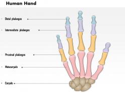 0514 the human hand medical images for powerpoint
We are proud to present our 0514 the human hand medical images for powerpoint. Anatomy of human palm is well explained in this medical power point diagram. Use this professional diagram in your presentation to explain human palm anatomy with bone structure study.
-
 0714 abdominal head of pectoralis major muscle medical images for powerpoint
We are proud to present our 0714 abdominal head of pectoralis major muscle medical images for powerpoint. This image contains the graphic of major chest muscles called as Pectoralis. We have used anterior view of abdominal head of pectoralis muscles to design this image. Use this image if you need to display human chest muscle in any presentation.
-
 0514 human eye anatomy medical images for powerpoint
We are proud to present our 0514 human eye anatomy medical images for powerpoint. The eye is a complex organ composed of many parts. Use this Medical Image to explain Structure of the Human Eye. The physical structure of the human eye enables it to sense light. This medical PowerPoint template can be used as communication tool for your medical presentations.
-
 0514 lateral cross sectional view of head and neck laryngeal anatomy medical images for powerpoint
We are proud to present our 0514 lateral cross sectional view of head and neck laryngeal anatomy medical images for powerpoint. This medical image helps in quickly and efficiently communicates your research. This allows viewers to study and restudy your information and discuss it with you one on one. You may use this medical image to give short presentations on your research.
-
 0514 anatomy of seif knee medical images for powerpoint
We are proud to present our 0514 anatomy of seif knee medical images for powerpoint. This Medical illustration depicts an anatomy of Seif Knee. One of the major joints in the body, the knee is required to support huge and repeated pressures over the course of a lifetime. Muscles keep bones in place and also play a role in movement of the bones. To allow motion, different bones are connected by joints.
-
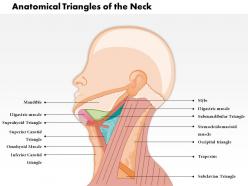 0514 anatomical triangles of neck medical images for powerpoint
We are proud to present our 0514 anatomical triangles of neck medical images for powerpoint. The anterior triangle of the neck is an anatomical division created by the muscles of the head and neck. It is used clinically to locate structures that pass through the neck. In this diagram take a look at the regional anatomy of the anterior triangle and its subdivisions.
-
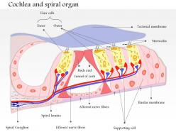 0514 cochlea and spiral organ medical images for powerpoint
We are proud to present our 0514 cochlea and spiral organ medical images for powerpoint. This Medical diagram Power Point template is designed with cochlea and spiral organ view in 3d. Use this template for explaining organs in any medical presentation.
-
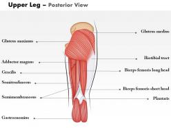 0514 upper legs posterior view medical images for powerpoint
We are proud to present our 0514 upper legs posterior view medical images for powerpoint. This Medical diagram template is designed with upper leg graphic with posterior view. Use it to explain human leg structure in your presentation and create an effect on your viewers.
-
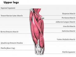 0514 upper legs anterior view medical images for powerpoint
We are proud to present our 0514 upper legs anterior view medical images for powerpoint. This Medical diagram template is designed with upper leg graphic with anterior view. Use it to explain human leg structure in your presentation and create an effect on your viewers.
-
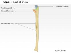 0514 ulna radial view medical images for powerpoint
We are proud to present our 0514 ulna radial view medical images for powerpoint. This Medical diagram template is designed with upper leg graphic with anterior view. Use it to explain human leg structure.
-
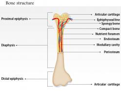 0714 bone structure medical images for powerpoint
We are proud to present our 0714 bone structure medical images for powerpoint. This image is crafted with bone structure. We have used internal view of human hand with various parts. Display proximal epiphysis, diaphysis and articular cartilage with this medical image. Make a professional presentation for your viewers with this suitable image.
-
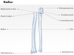 0514 radius ulnar view medical images for powerpoint
We are proud to present our 0514 radius ulnar view medical images for powerpoint. To explain bone structure you can use our great designer medical power point diagram in your presentations, this diagram is designed with ulnar view of radius bone.
-
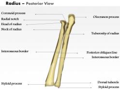 0514 radius posterior view medical images for powerpoint
We are proud to present our 0514 radius posterior view medical images for powerpoint. To explain bone structure you can use our great designer medical power point diagram in your presentations, this diagram is designed with posterior view of radius bone.
-
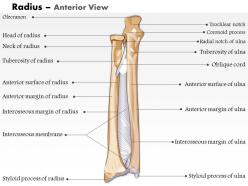 0514 radius anterior view medical images for powerpoint
We are proud to present our 0514 radius anterior view medical images for powerpoint. To explain bone structure you can use our great designer medical power point diagram in your presentations, this diagram is designed with anterior view of radius bone.
-
 0514 neck anterior view medical images for powerpoint
We are proud to present our 0514 neck anterior view medical images for powerpoint. To displaying body structure is very important for medical students. Use our medical diagrams in your presentation. This diagram template is designed with neck muscle graphic with anterior view ,use it to explain human neck structure.
-
 0514 muscles of neck medical images for powerpoint
We are proud to present our 0514 muscles of neck medical images for powerpoint. To displaying body structure is very important for medical students. Use our medical diagrams in your presentation. This diagram template is designed with neck muscle graphic ,use it to explain human neck structure.
-
 0514 femur posterior view medical images for powerpoint
We are proud to present our 0514 femur posterior view medical images for powerpoint. In medical science displaying body structure is very important and for that you can use our medical diagrams in your presentation. This diagram template is designed with femur bone graphic with posterior view. Use it to explain human leg structure.
-
 0514 female chest wall anterior view medical images for powerpoint
We are proud to present our 0514 female chest wall anterior view medical images for powerpoint. To display the anterior view of female chest use this medical diagram ppt. this medical diagram is designed with 3d graphic of female chest with anterior view. Use it to enhance the effect of your presentation.
-
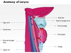 0514 anatomy of larynx medical images for powerpoint
We are proud to present our 0514 anatomy of larynx medical images for powerpoint. This Power Point template diagram is designed with 3d graphic of larynx. To describe the anatomy use this professional template in your presentation and give detail of larynx .
-
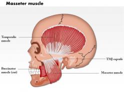 0714 masseter muscle medical images for powerpoint
We are proud to present our 0714 masseter muscle medical images for powerpoint. This medical image has been designed with internal view of masseter muscle. To explain this muscle, we have used graphic of human face with internal view. This muscle protects the jaw area. Use this image in your medical presentations.
-
 Human anatomy labelled bones skeleton
Presenting this set of slides with name Human Anatomy Labelled Bones Skeleton. This is a three stage process. The stages in this process are Human Anatomy Labelled Bones Skeleton. This is a completely editable PowerPoint presentation and is available for immediate download. Download now and impress your audience.
-
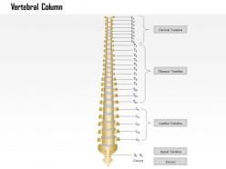 0914 vertebral column medical images for powerpoint
We are proud to present our 0914 vertebral column medical images for powerpoint. This medical image has been designed with graphic of Vertebral Column. This image is useful for nueuro and spine related presentations. Explain the Cervical, Vertebra and Thoracic in vertebral coloumn.
-
 0914 torn anterior cruciate ligament medical images for powerpoint
We are proud to present our 0914 torn anterior cruciate ligament medical images for powerpoint. This medicalimage has been crafted with human knee internal structure. This image explains the Anterior cruciate ligament ACL injuries in human knee. This image is useful for defining Femur, Normal Knee and Medial Collateral with Tibia in human knee.
-
 0914 infraspinatus muscle medical images for powerpoint
We are proud to present our 0914 infraspinatus muscle medical images for powerpoint. This medical image has been crafted with Infraspinatus Muscle. This image contains the human shoulder muscle with Supraspinatus, Teres Minor and Infraspinatus. Use this image for shoulder muscle related presentations.
-
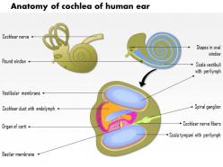 0914 anatomy of cochlea of human ear medical images for powerpoint
We are proud to present our 0914 anatomy of cochlea of human ear medical images for powerpoint. This medical image has been designed with graphic of human ear. This image contains the Anatomy Of Cochlea for Human Ear. Use this image to explain the internal cohlear nerve of human ear. Build presentations for internal view of human ear with all tagged parts.
-
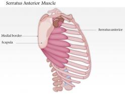 0714 serratus anterior muscle medical images for powerpoint
We are proud to present our 0714 serratus anterior muscle medical images for powerpoint. This image is a part of Musculoskeletal System. We have used graphic of serratus muscle with anterior view to display the details. This image contains parts of this muscle named as Medial border and Scapula. Use this image to explain the muscle in your medical presentations.
-
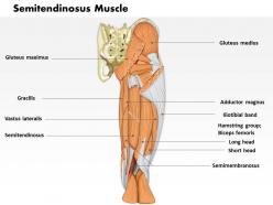 0714 semitendinosus muscle medical images for powerpoint
We are proud to present our 0714 semitendinosus muscle medical images for powerpoint. This medical image contains the graphic of semitendinosus muscle graphic. We have taken lateral view of this muscle with some internal parts. This muscle contains Gluteus medius Adductor Magnus Hamstring group Biceps femoris, which constitute this muscle. Create a stunning presentation with this professional image.
-
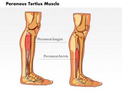 0714 peroneus tertius muscle medical images for powerpoint
We are proud to present our 0714 peroneus tertius muscle medical images for powerpoint. Peroneus muscle is present in human leg. To define the structure and importance of this muscle we have used interior view of human leg. In this image, we have defined Peroneus longus, Peroneus brevis and lower extensor retinaculum of this muscle. Use this image in your muscle related presentations and get good remarks.
-
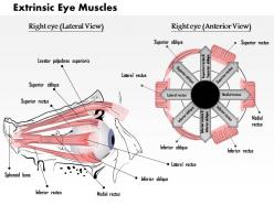 0514 the extrinsic eye muscles medical images for powerpoint
We are proud to present our 0514 the extrinsic eye muscles medical images for powerpoint. This Medical Power Point template is designed with the graphic of human eye with extrinsic muscle . To study the detail of human eye this view is very important use it in your presentation and make your views graphically strong.
-
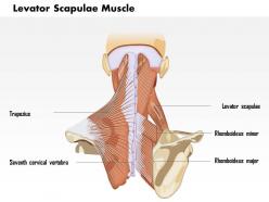 0714 levator scapulae muscle medical images for powerpoint
We are proud to present our 0714 levator scapulae muscle medical images for powerpoint. This quality medical image has been designed with external view of Levator scapulae muscle. This muscle is present on the neck area and is a protective layer of neck and backbone joint. This muscle contains Trapezius, Seventh cervical vertebra with Rhomboideus minor and major. Use this medical image to give excellent graphic viewed of muscle in any subject oriented presentations.
-
 0714 latissimus dorsi muscle medical images for powerpoint
We are proud to present our 0714 latissimus dorsi muscle medical images for powerpoint. This medical image is crafted with Latissimus muscle graphic. In human anatomy, this muscle is present in the chest area. This muscle contribute to protect ribs. Use this image in your presentations to explain Deltoid, Pectorals Major, Biceps Brachii and Latissimus Dorsi, which are the sub parts of this muscle.
-
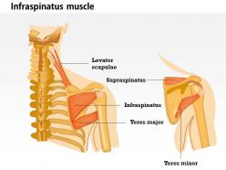 0714 infraspinatus muscle medical images for powerpoint
We are proud to present our 0714 infraspinatus muscle medical images for powerpoint. This medical image has been designed with infraspinatus muscle graphic. In human body, the infraspinatus muscle is a thick triangular muscle, which occupies the chief part of the infraspinatus fossa. Use this muscle graphic in your musculoskeletal presentations. You may also explain some of the parts of this muscle for example Levator scapulae, Supraspinatus, Infraspinatus and Teres major in your presentations.
-
 0714 human bony and muscular system posterior medical images for powerpoint
We are proud to present our 0714 human bony and muscular system posterior medical images for powerpoint. This medical image contains the human bone and muscular system graphic. In this image, we have taken posterior view of the system to give you a clear visibility. Explain parts of this muscle like Latissimus Dorsi, Posterior lilac Crest, Sacrum and Thoracolumbar Apo neurosis. Create a professional presentation for Musculoskeletal System using this image.
-
 0714 extensor carpi ulnaris muscle medical images for powerpoint
We are proud to present our 0714 extensor carpi ulnaris muscle medical images for powerpoint. To build a presentation for musculoskeletal system, use this medical image. In human anatomy, the extensor carpi ulnar is a skeletal muscle located on the ulnar side of the forearm. Add this image in your medical presentations and create an impact on viewers.
-
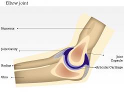 0714 elbow joint medical images for powerpoint
We are proud to present our 0714 elbow joint medical images for powerpoint. Display human elbow joint in any presentation with the help of this unique medical image. This image has been created with graphic of human elbow joint with anterior view. Explain the topic in a graphical way and make a good presentations for your audience.
-
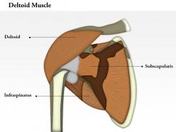 0714 deltoid muscle medical images for powerpoint
We are proud to present our 0714 deltoid muscle medical images for powerpoint. To display the graphic of human shoulder with internal structure, we have used highlighted part of muscle to give more effective representation.This image contains the graphic of one shoulder muscle, this muscle is called as deltoid. Explain the parts of this muscle like Sternum. Enhance the effect of your views with this innovative medical image.
-
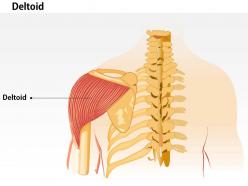 0714 deltoid medical images for powerpoint
We are proud to present our 0714 deltoid medical images for powerpoint. This image contains the graphic of one shoulder muscle, this muscle is called as deltoid. To display graphics of human shoulder with internal view, we have used highlighted part of the muscle to give it a more effective representation. Explain the parts of this muscle like Bicipital, Brachialis, Brachioradialis and Coracobrachialis. Use this fully editable image in your presentations and get attention of your viewers.
-
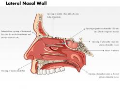 0514 lateral nasal wall medical images for powerpoint
We are proud to present our 0514 lateral nasal wall medical images for powerpoint. Medical Illustrations are especially useful in achieving what cameras cannot capture. In medical illustration, complex structures and concepts can be easily visualized. This medical illustration combines accuracy and clarity. Use this medical image to communicate effectively to a variety of audiences.
-
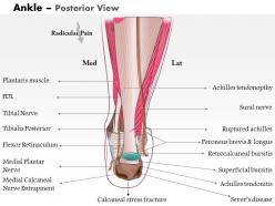 0514 ankle posterior medical images for powerpoint
We are proud to present our 0514 ankle posterior medical images for powerpoint. The above template shows Ankle Posterior View. Muscles of the ankle and foot function to move the ankle, foot, and toes and some are located in the lower leg. Fine muscles of ankle can exert tremendous power while constantly making small adjustments for balance.
-
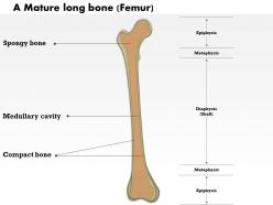 0514 a mature long bone medical images for powerpoint
We are proud to present our 0514 a mature long bone medical images for powerpoint. This medical diagram is designed with structure of long bone. Long bones are longer than they are wide and are the major bones of the limbs. Long bones grow more than the other classes of bone throughout childhood and so are responsible for the bulk of our height as adults.
-
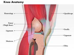 0514 side view of knee human anatomy medical images for powerpoint
We are proud to present our 0514 side view of knee human anatomy medical images for powerpoint. The knee joint is a relatively complex anatomical structure. During normal activity such as walking or running, and even for support while standing, the knee will function superbly. Use this medical image for full anatomical description of side view of human knee.
-
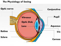 0514 physiology of seeing eye anatomy medical images for powerpoint
We are proud to present our 0514 physiology of seeing eye anatomy medical images for powerpoint. This PowerPoint template is an excellent tool for anyone who is learning basic eye anatomy and physiology. By using visuals such as medical images, concepts and ideas can be transferred quickly. For preparing an attractive and appealing presentation, you can add so many useful features in your display.
-
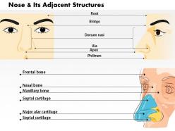 0514 nose and its adjacent structures medical images for powerpoint
We are proud to present our 0514 nose and its adjacent structures medical images for powerpoint. The above medical image is useful to understand the anatomy of the nose and its relationship to adjacent structures. This medical illustration combines accuracy and clarity. Use this readymade Medical PowerPoint template and save your time. This is the best way for presenting the ideas and concepts.
-
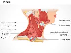 0514 neck medical images for powerpoint
We are proud to present our 0514 neck medical images for powerpoint. This template shows the neck medical image for PowerPoint. Use this interactive neck anatomy pictures for detailed descriptions. Use this medical image to communicate effectively to a variety of audiences. Medical illustration can be also be used to educate patients. Use this medical image to communicate effectively with them.
-
 0514 muscles of anterior neck and throat swallowing medical images for powerpoint
We are proud to present our 0514 muscles of anterior neck and throat swallowing medical images for powerpoint. This template shows the muscles of the anterior neck. Most of these muscles are deep muscles involved in swallowing. Swallowing begins when the tongue and buccinators muscles of the cheeks squeeze food. Use this template to give detailed presentation on muscles of anterior neck and throat swallowing.
-
 0514 lateral view of external nose anatomy of nasal skeleton medical images for powerpoint
We are proud to present our 0514 lateral view of external nose anatomy of nasal skeleton medical images for powerpoint. Use this interactive external nose anatomy pictures for descriptions of nasal skeleton. For preparing an attractive and appealing presentation, you can add this useful template in your display. Use this perfect medical template for appealing presentation and to hold the attention of the viewers.
-
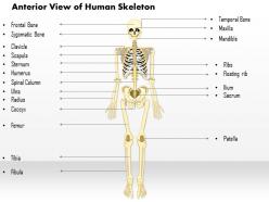 0514 anterior view of the human skeleton medical images for powerpoint
Useful for medical professionals and doctors operating on bones. No pixilation for graphics result in better projection on bigger or wider screens. PPT layout is convertible into JPEG and PDF and is easy to download and save. Layout does not occupy much of your disc space. Amendable font text, color scheme, orientation and design helps customize accordingly. Add company’s detail with respect to its identity with ease. No space constraints while mentioning titles and sub-titles.
-
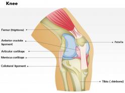 0514 knee medical images for powerpoint
We are proud to present our 0514 knee medical images for powerpoint. Use this medical image for full anatomical description of knee joint. Above PPT image shows different parts of the knee. Knee muscles are attached to two bones across a joint, so the muscle serves to move parts of those bones closer to each other. This medical PowerPoint template enables users to create elegant and appealing presentations that are able to attract the audience.
-
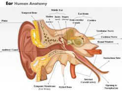 0514 ear human anatomy medical images for powerpoint
We are proud to present our 0514 ear human anatomy medical images for powerpoint. This Medical illustration will help you to understand ear human anatomy. In medical illustration, complex structures and concepts can be easily visualized. This diagram of the human ear helps in visual interpretations or the structure and function of the human ear. This template is being designed properly to attract the users attention.
-
 0514 cartilaginous and bony structures of nasal septum medical images for powerpoint
We are proud to present our 0514 cartilaginous and bony structures of nasal septum medical images for powerpoint. Above shown nasal septum PowerPoint diagram is designed with scientific accuracy. By using this medical illustration concepts and ideas can be transferred quickly. This is a product of concise communication for a medical illustrator to reviewing the anatomy. This template is being designed properly to attracts the users attention.
-
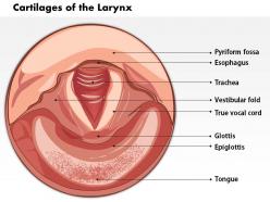 0514 cartilages of the larynx medical images for powerpoint
We are proud to present our 0514 cartilages of the larynx medical images for powerpoint. This image shows the cartilages of the larynx. The Larynx serves a number of purposes. It is designed specifically for our speaking and singing, the larynx has evolved to allow us this control. It has other biological purposes too, ones that are essential to life.
-
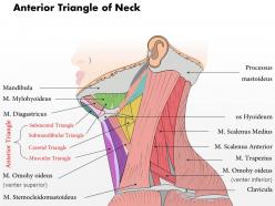 0514 anterior triangle of neck medical images for powerpoint
We are proud to present our 0514 anterior triangle of neck medical images for powerpoint. The anterior triangle of the neck is an anatomical division created by the muscles of the head and neck. It is used clinically to locate structures that pass through the neck. Use this diagram to have a look at the anatomy of the anterior triangle and its subdivisions.
-
 0514 anatomy of knee joint medical images for powerpoint
We are proud to present our 0514 anatomy of knee joint medical images for powerpoint. Use this medical image for full anatomical description of knee joint. Above PPT image shows different parts of the knee. Knee muscles are attached to two bones across a joint, so the muscle serves to move parts of those bones closer to each other. This medical PowerPoint template enables users to create elegant and appealing presentations that are able to attract the audience.
-
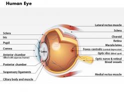 0514 anatomy of human eye medical images for powerpoint
We are proud to present our 0514 anatomy of human eye medical images for powerpoint. The eye is a complex organ composed of many parts. Use this Medical Image to explain Structure of the Human Eye. The physical structure of the human eye enables it to sense light. This medical PowerPoint template can be used as communication tool for your medical presentations.
-
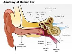 0514 anatomy of human ear medical images for powerpoint
We are proud to present our 0514 anatomy of human ear medical images for powerpoint. Use this PPT diagram to explain how the human ear serves as an astounding transducer, converting sound energy to mechanical energy to a nerve impulse that is transmitted to the brain. Anatomy of human ear shows how humans hear is a complex subject involving the fields of physiology.
-
 0514 ulna posterior medical images for powerpoint
We are proud to present our 0514 ulna posterior medical images for powerpoint. The above template shows Ulna posterior view Medical Images for PowerPoint. The ulna is one of two bones that make up the forearm. The ulna is located on the opposite side of the forearm from the thumb,
-
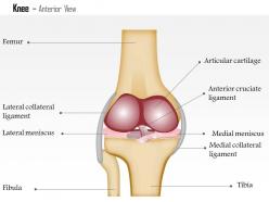 0514 knee anterior view medical images for powerpoint
We are proud to present our 0514 knee anterior view medical images for powerpoint. This Medical Power Point template is designed with knee anterior view for human anatomy related presentation. Use this template to impress your seniors or to give better explanation of the topic to your juniors.
-
 0514 human foot medical images for powerpoint
We are proud to present our 0514 human foot medical images for powerpoint. This Medical Power Point template is designed with the graphic of human foot in plantar condition. To study the detail of human foot this view is very important use it in your presentation and make your views graphically strong.
-
 0514 human anatomy elbow anterior view medical images for powerpoint
We are proud to present our 0514 human anatomy elbow anterior view medical images for powerpoint. Graphic of human elbow is used in this Medical Power Point template to define human elbow anatomy structure. Use this template for explaining the elbow structure in your anatomy presentation.
-
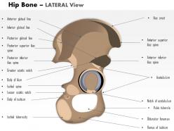 0514 hip bone lateral view medical images for powerpoint
We are proud to present our 0514 hip bone lateral view medical images for powerpoint. This Medical Power Point template is designed with lateral view of human hip bone. Explain internal structure of human hip bone with our professional diagram template in your educational presentation.
-
Great experience, I would definitely use your services further.
-
Design layout is very impressive.
-
Design layout is very impressive.
-
Best way of representation of the topic.
-
Excellent design and quick turnaround.
-
Informative presentations that are easily editable.
-
Great quality product.
-
I discovered this website through a google search, the services matched my needs perfectly and the pricing was very reasonable. I was thrilled with the product and the customer service. I will definitely use their slides again for my presentations and recommend them to other colleagues.
-
Best Representation of topics, really appreciable.
-
Excellent products for quick understanding.


